The heart is one of the most important organs in the human body. It works like a powerful pump that keeps blood circulating throughout the body, delivering oxygen and nutrients while removing waste products. To do this efficiently, the heart is made up of several important parts, each with a specific role. In this article, we’ll explore the different parts of the heart and understand how they work together.
List Of Heart Parts With Names
- Aorta
- Superior vena cava
- Right pulmonary artery
- Pulmonary trunk
- Right pulmonary veins
- Right atrium
- Fossa ovalis
- Tricuspid valve
- Right ventricle
- Chordae tendineae
- Trabeculae carneae
- Moderator band
- Inferior vena cava
- Left pulmonary artery
- Left atrium
- Left pulmonary veins
- Mitral (bicuspid) valve
- Aortic valve
- Pulmonary valve
- Left ventricle
- Papillary muscle
- Interventricular septum
- Epicardium
- Myocardium
- Endocardium
Parts of the Heart Diagram With Names
The heart is a vital organ that works as a powerful pump to circulate blood throughout the body. It is made up of several important parts that work together to maintain proper blood flow. The chambers of the heart receive and pump blood, while the valves ensure that blood flows in the correct direction without flowing backward. Major blood vessels carry blood to and from the heart, connecting it with the lungs and the rest of the body. In addition, the heart wall is made of special layers and muscles that help it contract and relax with every heartbeat. Each part, from the atria and ventricles to the valves and vessels, plays an essential role in keeping the circulatory system working efficiently.
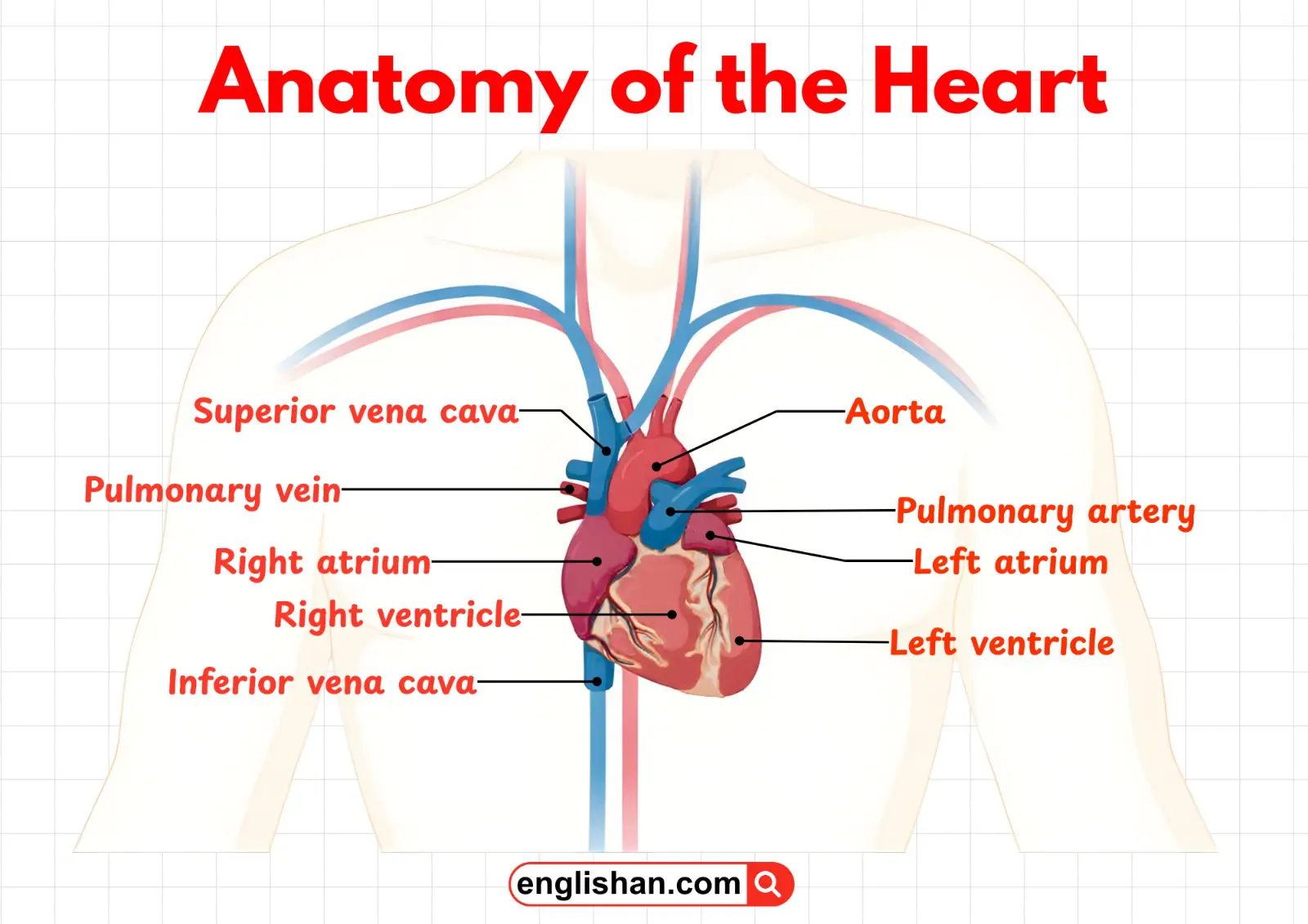
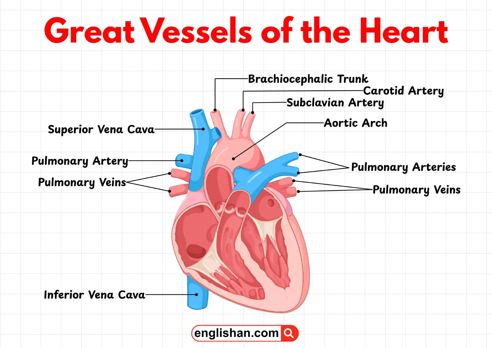
Major Blood Vessels
Aorta
The aorta is the largest artery in the human body. It carries oxygen-rich blood from the left ventricle of the heart to the rest of the body. After blood leaves the heart through the aortic valve, it enters the aorta and is distributed through a network of arteries to supply oxygen and nutrients to tissues and organs.
Superior Vena Cava
The superior vena cava is a large vein that brings deoxygenated blood from the upper part of the body, including the head, neck, arms, and chest, back to the heart. This blood enters the right atrium where it will later be sent to the lungs for oxygenation.
Inferior Vena Cava
The inferior vena cava is the largest vein in the lower part of the body. It carries deoxygenated blood from the lower body, including the abdomen, pelvis, and legs, to the right atrium of the heart. It works together with the superior vena cava to return blood to the heart.
Pulmonary Trunk
The pulmonary trunk is a large blood vessel that carries deoxygenated blood from the right ventricle to the lungs. Shortly after leaving the heart, it divides into the left and right pulmonary arteries that transport blood to each lung for oxygenation.
Right Pulmonary Artery
The right pulmonary artery carries deoxygenated blood from the pulmonary trunk to the right lung. In the lungs, the blood releases carbon dioxide and receives oxygen before returning to the heart.
Left Pulmonary Artery
The left pulmonary artery carries deoxygenated blood from the pulmonary trunk to the left lung. Like the right pulmonary artery, it allows the blood to undergo gas exchange in the lungs
Right Pulmonary Veins
The right pulmonary veins carry oxygenated blood from the right lung back to the left atrium of the heart. Unlike most veins, pulmonary veins carry oxygen-rich blood.
Left Pulmonary Veins
The left pulmonary veins transport oxygenated blood from the left lung to the left atrium. These veins complete the pulmonary circulation by returning freshly oxygenated blood to the heart.
Heart Chambers
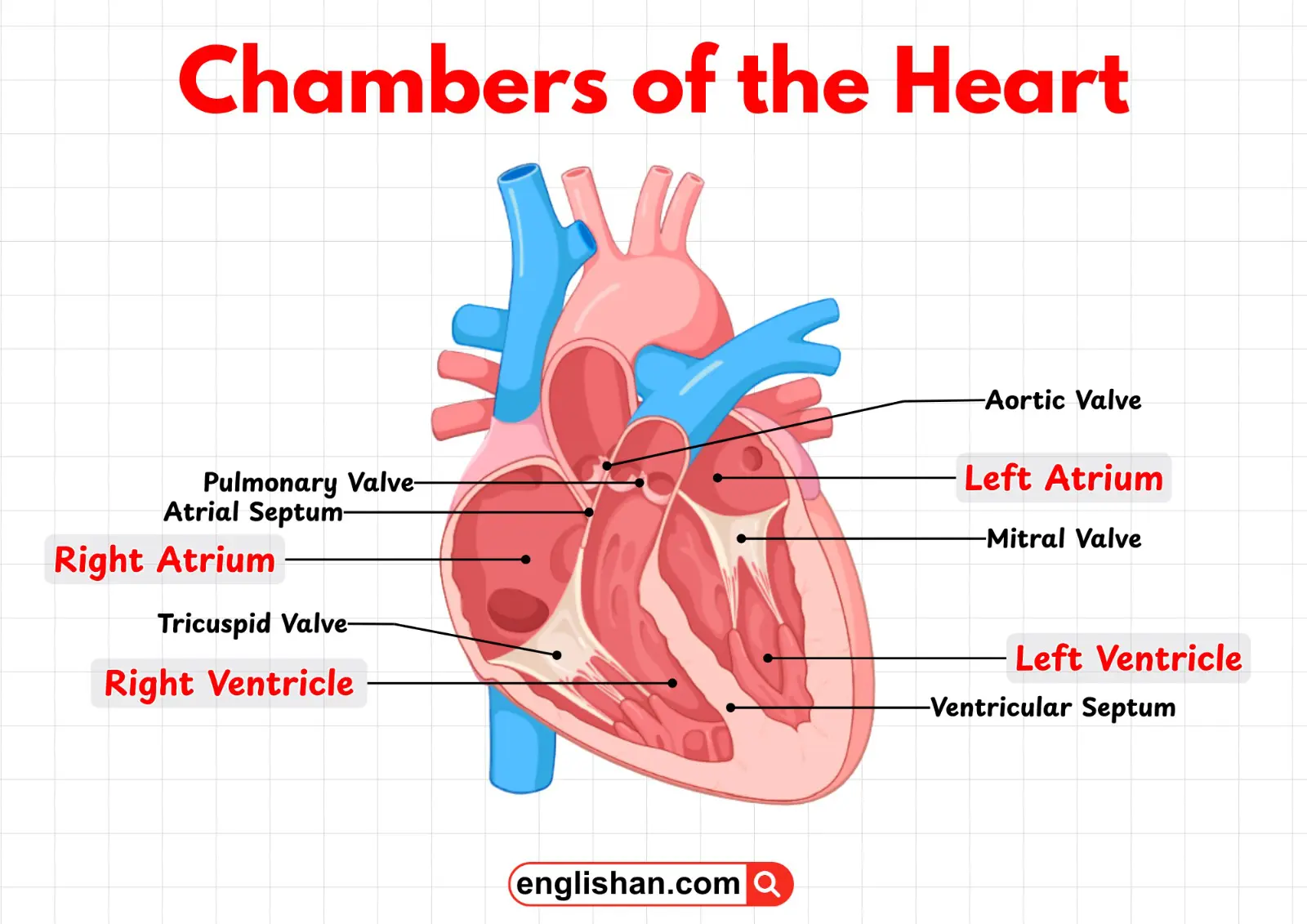
Right Atrium
The right atrium is the upper right chamber of the heart. It receives deoxygenated blood from the superior vena cava and inferior vena cava. This blood is then passed through the tricuspid valve into the right ventricle.
Left Atrium
The left atrium is the upper left chamber of the heart. It receives oxygenated blood from the lungs through the pulmonary veins. The blood then moves through the mitral valve into the left ventricle.
Right Ventricle
The right ventricle is the lower right chamber of the heart. It pumps deoxygenated blood to the lungs through the pulmonary valve and pulmonary trunk for oxygenation.
Left Ventricle
The left ventricle is the strongest and most muscular chamber of the heart. It pumps oxygenated blood through the aortic valve into the aorta so that it can be distributed throughout the body.
Heart Valves
Tricuspid Valve
The tricuspid valve is located between the right atrium and the right ventricle. It has three flaps (cusps) and ensures that blood flows in one direction—from the right atrium to the right ventricle—without flowing backward.
Mitral (Bicuspid) Valve
The mitral valve lies between the left atrium and the left ventricle. It has two flaps and controls blood flow from the left atrium to the left ventricle, preventing backflow during ventricular contraction.
Pulmonary Valve
The pulmonary valve is located between the right ventricle and the pulmonary trunk. It opens to allow blood to flow toward the lungs and closes to prevent blood from flowing back into the right ventricle.
Aortic Valve
The aortic valve is located between the left ventricle and the aorta. It opens when the left ventricle contracts to allow blood to enter the aorta and closes to prevent blood from returning to the heart.
Internal Muscles & Structural Components
Papillary Muscle
Papillary muscles are small muscles located inside the ventricles of the heart. They attach to the chordae tendineae and contract during heartbeats to prevent the valves from turning inside out when the ventricles contract.
Chordae Tendineae
Chordae tendineae are thin, strong, tendon-like cords that connect the heart valves to the papillary muscles. They stabilize the valves and prevent them from collapsing backward during ventricular contraction.
Trabeculae Carneae
Trabeculae carneae are irregular muscular ridges found on the inner walls of the ventricles. These ridges help the heart contract more efficiently and prevent suction that could interfere with blood flow.
Moderator Band
The moderator band is a muscular band found in the right ventricle. It helps carry electrical signals through the heart muscle and assists in coordinating the contraction of the right ventricle.
Interventricular Septum
The interventricular septum is the thick muscular wall that separates the right and left ventricles. It prevents oxygenated and deoxygenated blood from mixing and also plays a role in the heart’s electrical conduction system.
Fossa Ovalis
The fossa ovalis is a small depression in the wall between the right and left atria. It is a remnant of the foramen ovale, an opening that existed in the fetal heart to allow blood to bypass the lungs before birth.
Layers of the Heart Wall
Epicardium
The epicardium is the outermost layer of the heart wall. It protects the heart and forms part of the protective sac surrounding the heart known as the pericardium.
Myocardium
The myocardium is the thick, muscular middle layer of the heart wall. It is responsible for the pumping action of the heart, as its muscle fibers contract to push blood through the circulatory system.
Endocardium
The endocardium is the thin inner lining of the heart chambers and valves. It provides a smooth surface that allows blood to flow easily through the heart without friction.
Key Takeaway
As we learned, the heart is a complex organ made up of several important parts that work together to keep blood circulating throughout the body. The chambers receive and pump blood, the valves ensure one-way blood flow, and the blood vessels transport blood to and from the heart. In addition, the muscles and layers of the heart help it contract and relax efficiently with every heartbeat. Together, all these parts play a crucial role in maintaining a healthy and properly functioning circulatory system.
You May Also Like