In this blog post, you will learn the different parts of the foot in English. The foot is an important part of the body that helps with movement and balance. Knowing the correct names of its parts can improve your vocabulary and understanding of body-related terms. This knowledge is useful for everyday conversations, medical discussions, and learning human anatomy in English.
To learn more vocabulary on different topics, visit our Vocabulary Category.
Regions of Foot
The foot can be divided into several regions, each with specific structures and functions. Here are the main regions of the foot:
1. Rearfoot:
- Calcaneus (Heel Bone): The large bone at the back of the foot, providing a foundation for weight-bearing and stability.
- Talus: The bone that forms the ankle joint with the tibia and fibula.
2. Midfoot:
- Navicular: A boat-shaped bone located in the arch of the foot.
- Cuboid: A cube-shaped bone on the outer side of the foot.
- Cuneiform Bones (Medial, Intermediate, Lateral): Three wedge-shaped bones forming the arch of the foot.
3. Forefoot:
- Metatarsals (1-5): Long bones extending from the midfoot to the toes.
- Phalanges (Proximal, Middle, Distal): Toe bones, with each toe typically having three phalanges except for the big toe, which has two.
4. Sole (Plantar Surface):
- The bottom surface of the foot that contacts the ground, including the:
- Plantar fascia: A thick band of tissue supporting the arch.
- Fat pads: Providing cushioning and shock absorption.
5. Dorsum (Top of the Foot):
- The upper surface of the foot, including tendons and blood vessels.
6. Ankle:
- The joint connecting the foot to the leg, consisting of the tibia, fibula, and talus bones.
7. Toes:
- The front part of the foot, comprising the toes (phalanges) and metatarsophalangeal joints.
8. Arch Structures:
- Medial Longitudinal Arch: Along the inner side of the foot.
- Lateral Longitudinal Arch: Along the outer side of the foot.
- Transverse Arch: Running across the foot.
Bones and Joints
Bones of the Foot:
1. Calcaneus (Heel Bone):
Located at the back of the foot, providing a stable base for body weight.
2. Talus:
Articulates with the tibia and fibula to form the ankle joint, allowing up-and-down movement.
3. Navicular:
Positioned in the midfoot, contributing to the foot’s arches.
4. Cuboid:
Found on the outer side of the foot, connecting with the metatarsals.
5. Cuneiform Bones (Medial, Intermediate, Lateral):
Wedge-shaped bones in the midfoot, supporting the arch.
6. Metatarsals (1-5):
Long bones extending from the midfoot to the toes.
7. Phalanges (Proximal, Middle, Distal):
Toe bones, with each toe typically having three phalanges except for the big toe, which has two.
Joints of the Foot:
1. Ankle Joint:
Formed by the tibia, fibula, and talus bones, allowing up-and-down movement.
2. Subtalar Joint:
Between the talus and calcaneus, enabling side-to-side movement.
3. Midfoot Joints:
Various joints between the tarsal bones (navicular, cuboid, and cuneiforms), contributing to flexibility and weight distribution.
4. Metatarsophalangeal Joints:
Connect the metatarsals to the proximal phalanges, allowing movement of the toes.
5. Interphalangeal Joints:
Joints between the phalanges, facilitating bending and straightening of the toes.
The tibia and fibula are two long bones located in the lower leg, forming an essential part of the human skeletal system.
Anatomy
Foot anatomy refers to the complex structure of the human foot, a remarkable body part designed for weight-bearing, balance, and locomotion. The foot comprises numerous bones, joints, muscles, tendons, ligaments, and arches, all working together to support the body and facilitate various movements. The main components of foot anatomy include:
1. Bones:
The foot has 26 bones, including the tarsal bones (such as the calcaneus, talus, navicular, cuboid, and cuneiforms), metatarsals (long bones in the midfoot), and phalanges (toe bones).
2. Joints:
Key joints include the ankle joint (formed by the tibia, fibula, and talus), subtalar joint (between the talus and calcaneus), and various metatarsophalangeal and interphalangeal joints.
3. Muscles:
Intrinsic muscles are located within the foot and control fine movements, while extrinsic muscles extend from the leg and control larger movements.
4. Tendons:
Tendons connect muscles to bones and play a crucial role in transmitting forces during movement.
5. Ligaments:
Ligaments connect bones to bones, providing stability to the joints of the foot.
6. Arches:
The foot has three arches—the medial longitudinal arch, lateral longitudinal arch, and transverse arch—that contribute to weight distribution, shock absorption, and adaptability to different surfaces.
Structure of Foot
The foot is a complex anatomical structure with various bones, joints, muscles, ligaments, tendons, and other tissues working together to support the body and enable movement. Here is an overview of the structure of the foot:
1. Bones:
The foot is composed of 26 bones, which can be grouped into three main sections:
- Hindfoot: Includes the calcaneus (heel bone) and the talus.
- Midfoot: Comprises the navicular, cuboid, and cuneiform bones (medial, intermediate, and lateral).
- Forefoot: Contains the metatarsals (long bones in the midfoot) and the phalanges (toe bones).
2. Joints:
- Ankle Joint: Formed by the tibia, fibula, and talus bones, allowing up-and-down movement.
- Subtalar Joint: Between the talus and calcaneus, enabling side-to-side movement.
- Midfoot Joints: Various joints among the tarsal bones, contributing to flexibility.
- Metatarsophalangeal Joints: Connect the metatarsals to the proximal phalanges.
- Interphalangeal Joints: Joints between the phalanges.
Muscles:
- Intrinsic muscles are located within the foot, controlling fine movements.
- Extrinsic muscles, originating outside the foot, control larger movements and extend into the foot through tendons.
Ligaments:
- Ligaments connect bones to bones, providing stability to the joints. Examples include the plantar fascia and various ankle ligaments.
Tendons:
- Tendons connect muscles to bones, facilitating movement. The Achilles tendon is a notable example.
Arches:
The foot has three arches:
- Medial Longitudinal Arch: Along the inner side of the foot.
- Lateral Longitudinal Arch: Along the outer side of the foot.
- Transverse Arch: Running across the foot.
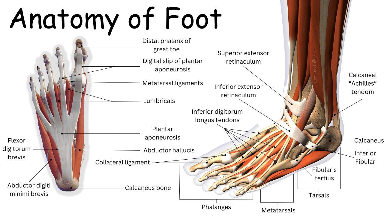
Anatomy of Foot
The anatomy of the foot is a complex and highly specialized structure designed to support the body, provide stability, and facilitate various movements. Here is a breakdown of the key components of foot anatomy:
Calcaneal Tendon:
The strong band that connects your calf muscles to your heel bone, also known as the Achilles tendon.
Calcaneus:
The big bone at the back of your foot, right under your ankle. It’s like your foot’s foundation.
Inferior Fibular:
The lower end of the smaller bone in your lower leg, the one on the outer side.
Fibularis Tertius:
A muscle on the front of your lower leg that helps lift your foot and turn it outward.
Tarsals:
These are the seven small bones in the middle of your foot, including your heel bone and the ones leading up to your toes.
Metatarsals:
The longer bones in your foot, right between your tarsals and your toe bones.
Collateral Ligament:
Think of this like a little band on the sides of your toes that helps keep them steady.
Phalanges:
Fancy word for the bones in your toes – you’ve got three in each toe.
Flexor Digitorum Brevis:
A little muscle in your foot that helps your toes bend.
Abductor Digiti Minimi Brevis:
Another tiny muscle, this one helps move your pinky toe away from the others.
Distal Phalanx of Great Toe:
The bone at the very end of your big toe.
Digital Slip of Plantar Aponeurosis:
This is like a stretchy band in the bottom of your foot that helps keep your arch up.
Metatarsal Ligaments:
Little bands that give support to the bones in the middle of your foot.
Lumbricals:
These are special muscles that help your toes bend and straighten.
Calcaneus Bone:
The big bone that makes up your heel, helping you stand and balance.
Common Foot Problems
Several common foot problems can affect people of all ages, often causing discomfort and affecting mobility. Here are some of the most prevalent foot issues:
- Athlete’s Foot: A fungal infection causing itching, redness, and cracked skin, usually between the toes.
- Plantar Fasciitis: Inflammation of the plantar fascia, a band of tissue along the bottom of the foot, leading to heel pain, especially in the morning.
- Bunions: A bony bump at the base of the big toe, causing the toe to angle towards the other toes.
- Ingrown Toenails: Toenails that grow into the surrounding skin, often causing pain, swelling, and infection.
- Corns and Calluses: Thickened and hardened areas of skin, typically on the toes or soles, due to friction or pressure.
- Hammer Toe: Abnormal bending of the toe joints, causing them to resemble a hammer.
- Flat Feet: A condition where the arches of the feet collapse, leading to a flat appearance and sometimes causing pain.
- Heel Spurs: Bony outgrowths on the heel bone, often associated with plantar fasciitis and causing heel pain.
- Achilles Tendonitis: Inflammation of the Achilles tendon, resulting in pain and stiffness at the back of the heel.
- Neuromas: Thickening of nerve tissue between the toes, causing pain and discomfort.
- Gout: A form of arthritis characterized by sudden, severe attacks of pain, swelling, and redness, often affecting the big toe.
- Morton’s Neuroma: Thickening of tissue around a nerve in the ball of the foot, leading to pain, tingling, and numbness.
- Fungal Nail Infections: Infections of the toenails caused by fungi, resulting in discoloration, thickening, and crumbling of the nails.
- Sprains and Strains: Overstretching or tearing of ligaments (sprains) or muscles/tendons (strains) in the foot, often caused by injury or overuse.
- Tendonitis: Inflammation of tendons, leading to pain and swelling, often occurring in the Achilles or other foot tendons.
Medical Conditions
Medical conditions affecting the feet can vary widely, ranging from common issues to more complex and systemic conditions. Here is a list of medical conditions that can impact the feet:
Diabetes-related Foot Issues:
- Peripheral Neuropathy: Nerve damage causing numbness, tingling, and pain.
- Foot Ulcers: Open sores that may develop due to reduced sensation and circulation.
Arthritis:
- Osteoarthritis: Degeneration of joint cartilage, often affecting the big toe and midfoot.
- Rheumatoid Arthritis: Autoimmune disorder causing joint inflammation, impacting foot joints.
Peripheral Arterial Disease (PAD):
- Reduced blood flow to the extremities, leading to cramping, pain, and slow wound healing.
Gout:
- Buildup of uric acid crystals in the joints, commonly affecting the big toe.
Circulatory Disorders:
- Deep Vein Thrombosis (DVT): Blood clot formation in deep veins, potentially affecting blood flow to the feet.
- Raynaud’s Disease: Reduced blood flow to extremities, causing numbness and discoloration.
Neuromuscular Disorders:
- Charcot-Marie-Tooth Disease (CMT): Inherited neurological disorder affecting the nerves in the feet and legs.
- Multiple Sclerosis (MS): Autoimmune disorder affecting the central nervous system, potentially causing foot numbness and weakness.
Psoriasis and Psoriatic Arthritis:
- Skin condition (psoriasis) and associated arthritis affecting joints, including those in the feet.
Peripheral Neuropathy:
- Nerve damage leading to pain, tingling, or loss of sensation, often associated with conditions like diabetes.
Buerger’s Disease:
- Inflammatory condition affecting blood vessels, particularly in the feet and hands.
Corns and Calluses:
- Thickened skin caused by friction or pressure, often linked to improper footwear.
Charcot Foot:
- Weakening of bones in the foot due to nerve damage, often seen in individuals with diabetes.
Osteoporosis:
- Decreased bone density, increasing the risk of fractures in the feet.
Foot Care
Proper Footwear Selection:
- Choosing shoes with proper support and fit.
Daily Foot Hygiene:
- Washing, drying, and inspecting feet regularly.
Prompt Pain Addressing:
- Seeking attention for persistent foot pain.
Regular Foot Inspections:
- Checking for cuts, blisters, and changes in skin color.
Correct Toenail Trimming:
- Trimming toenails straight across, avoiding short cuts.
Ingrown Toenail Awareness:
- Not ignoring signs and seeking prompt attention.
Sunscreen Application:
- Protecting feet from sun exposure.
Moisturization Routine:
- Keeping feet moisturized while avoiding areas between toes.
Safe Callus Care:
- Using gentle methods to address calluses.
Diabetes Foot Care:
- Following a strict foot care routine for diabetes management.
Regular Foot Stretches:
- Incorporating stretches for flexibility.
Routine Exercise:
- Engaging in regular physical activity for circulation.
Understanding Foot Anatomy:
- Making informed choices based on foot structure.
Scheduled Professional Check-ups:
- Regular visits to a podiatrist or healthcare professional.
Caution with Self-Treatment:
- Avoiding self-diagnosis and treatment for serious conditions.
Avoiding Mistakes
You May Also Like


